Breast Thermography
Thermal Imaging for Overall Breast Health — safe, non-invasive, and radiation-free early detection for women of all ages.
Book Your Scan
Pricing & Booking
Your thermogram includes detailed thermal imaging using FDA-inspected equipment. Board-certified thermologists interpret your results. All pricing is upfront — no hidden fees.
Breast Baseline Package
- Initial Breast Thermography Scan
- 3-Month Comparison Scan
- Doctor-Interpreted Reports for Both Visits
Two scans 90 days apart establish your personal thermal baseline for all future monitoring.
Breast Initial Scan
- Breast & Chest
- Side angles included
- Full MD interpretation
First-time patients require two scans 90 days apart to establish a baseline. We recommend the package for best value.
Breast Follow-Up Scan
- Breast & Chest
- Side angles included
- Compared to your baseline
For returning patients with an established baseline. Annual follow-up recommended.
No Referral Needed
Empowering women to manage their breast health independently — no referral or insurance approval required.
Radiation-Free & Non-Invasive
A safe, holistic option for those seeking alternatives to traditional invasive screenings.
Medical-Grade Equipment
FDA-inspected medical-grade infrared imaging for unparalleled precision in every scan.
Comfortable & Quick Session
Conducted by a board-certified thermographer in a supportive, professional environment.
Board Certified MD Analysis
Images analyzed by board-certified thermologists. Full medical interpretation included — no surprise fees.
HIPAA Compliant Report
Digital report shared with you and any providers you choose — at no additional cost.
* First-time breast scans require two scans for effectiveness — an initial scan and a follow-up 90 days later to establish your personal thermal baseline. Save $61 by booking the Breast Package bundle.
"1 in 8 women in the United States will be diagnosed with breast cancer in her lifetime." Source: National Breast Cancer Foundation Inc.

"When caught in its earliest, localized stages, the 5-year relative survival rate is 99%." Source: National Breast Cancer Foundation Inc.

Detect Disease at the Earliest Stage
Thermography offers the opportunity of earlier detection of breast disease than has been possible through breast self-examination or mammography alone. It captures infrared radiation from the skin and translates it into a visual color-spectrum map — identifying temperature variations as small as 1/100th of a degree, indicative of increased blood supply and chemical activity often seen in developing breast pathology.
Average Growth Rate of a Breast Cancer Tumor — cells double approximately every 90 days
Thermography Detects Irregularities At Any Stage
Thermography can identify areas of thermal asymmetry early in cell development — findings that, when followed up promptly with your physician, may lead to earlier diagnostic testing and more treatment options.
Mammogram Detection Begins at 1cm+
Mammograms detect breast cancer only when cells form a detectable mass — typically at least 1cm — indicating cells have already doubled 32 times.*
*Source: Buchanan JB, et al. Tumor growth, doubling times, and inability of the radiologist to diagnose certain cancers. Radiol Clin N Am. 1983;21:115-26

Understanding the Heat Signature
In areas where breast pathology is developing, the activity of chemicals and blood vessels is often much higher than in normal breast tissue. Cancer cells require a substantial supply of nutrients to grow — a process known as angiogenesis — which raises the surface temperature of the breast. Thermography uniquely measures this increase in heat.
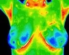
A "normal" scan exhibits balanced symmetry without significant hot or cool spots, serving as a reference point for future breast thermograms.
Fibrocystic scans typically appear as mildly warm areas on a thermal scan and are assessed by comparison to a second baseline adjusted for age.
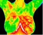
Vascular asymmetry led to clinical investigation revealing a palpable mass. A biopsy verified DCIS (Ductal Carcinoma In Situ), and the patient underwent surgery to have the tumor excised.

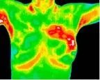
This cancer type is undetectable by mammography as it does not form a "lump." A biopsy recommended after thermography diagnosed inflammatory cancer at a very early stage.
Thermography vs. Mammogram vs. Ultrasound
All three are valuable breast health tools — each with distinct strengths. Understanding the differences helps you build the most complete picture of your breast health.
| Thermography | Mammogram | Ultrasound | |
|---|---|---|---|
| What it is | Infrared imaging that detects heat and vascular changes in tissue — a test of physiology and function, not structure. | An X-ray that produces structural images of breast tissue to identify masses, calcifications, and densities. | Sound waves that produce real-time structural images — used to characterize masses found on mammogram or exam. |
| Purpose | Detect thermovascular and physiological changes that can precede structural changes by years — focused on early risk detection. | Detect existing structural masses and calcifications in breast tissue — primarily for women 40+ or those with symptoms. | Clarify or characterize findings from mammogram or physical exam — particularly useful for distinguishing cysts from solid masses. |
| Radiation | None — completely radiation-free. Safe for women of all ages including younger women and those avoiding radiation exposure. | Low-dose ionizing radiation (X-ray). Cumulative exposure from annual screening is considered low-risk by most guidelines. | None — ultrasound uses sound waves, not radiation. Safe for all ages. |
| Comfort | No contact, no compression, no discomfort. Patient simply stands before the camera in a temperature-controlled room. | Requires breast compression, which many women find uncomfortable or painful. Compression is necessary for image clarity. | Requires gel application and handheld probe contact. Generally comfortable but involves direct contact with the breast. |
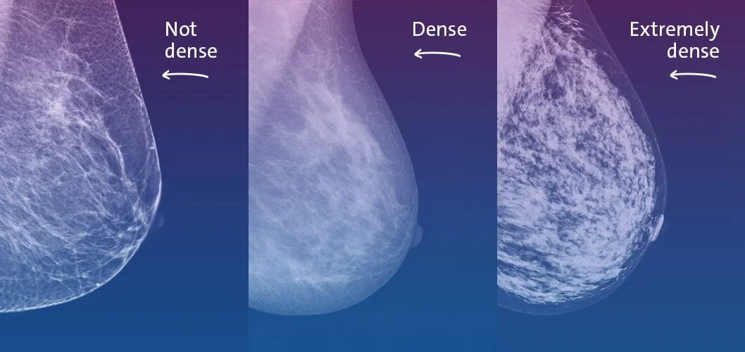
| Dense Breasts | Unaffected by tissue density. Measures heat and vascular activity — not tissue structure — making it equally effective regardless of density. | Significantly limited by dense tissue. Studies show mammograms miss up to 50% of cancers in women with dense breasts.* | Better than mammogram for dense breasts — often used as a supplement. Still limited to structural findings only. |
| Detection Type | Detects physiological and vascular changes (neoangiogenesis) that may occur before a mass becomes structurally present.* | Detects structural masses typically ≥1cm — meaning cancer cells have already doubled approximately 32 times before detection.* | Detects existing structural masses — similar timeline to mammogram. Does not detect pre-structural physiological changes. |
| Age Suitability | Appropriate for women of all ages — including women under 40 who are not yet recommended for routine mammography. | Routine screening typically recommended from age 40–50 depending on guidelines. Limited utility for younger women. | Used at any age when clinically indicated — typically following a mammogram finding or palpable lump, not as routine screening. |
| FDA Status | FDA-cleared as an adjunct screening tool — intended to complement, not replace, other breast health imaging. | FDA-approved as a primary breast cancer screening tool. | FDA-cleared as a diagnostic and adjunct imaging tool — not approved as a standalone screening method. |
| Bottom Line | The earliest physiological risk detection available — measures what's happening in tissue before structure changes. Best used alongside mammogram and/or ultrasound. | The gold standard for structural breast cancer screening — best for detecting existing masses and calcifications in women 40+. | A valuable complement to mammogram for characterizing masses — particularly effective in dense breast tissue. Not a standalone screening tool. |
The expert consensus: Thermography, mammography, and ultrasound are complementary — not competing — tools. Thermography excels at detecting physiological risk early; mammogram and ultrasound excel at identifying and characterizing existing structural findings.
* Sources: Buchanan JB et al., Radiol Clin N Am. 1983; National Cancer Institute; Amalu WC, International Academy of Clinical Thermology.
Early Detection Guidelines
Thermography and mammography are not in competition but rather complement each other. The aim is always the earliest possible intervention, leveraging the strengths of multiple screening methods.
- Annual breast examination by your physician.
- Annual breast thermography screening for women of all ages.
- Mammography, when considered appropriate for women who are aged 50 or older and/or recommended by your physician.
- Personal awareness for changes in the breasts by monthly self breast examination.
- Readiness to discuss quickly any such changes with a doctor.
Thermography Fills Gaps in Early Detection
Thermography stands out as a vital early detection tool for women under 40, women with dense breasts, and those at risk of Inflammatory Breast Disease — areas where mammograms often fall short.
Breast cancers grow faster in women under age 50 — Source: Cancer 71:3547-3551, 1993
Early detection options for younger women.
Breast thermography particularly benefits younger women aged 30-50, whose denser breast tissue may render mammography less effective. Breast cancer is the most common cancer among women aged 15-39 — a demographic not typically recommended for mammograms.

| Age | Average Doubling Time |
|---|---|
| Under 50 | 80 Days |
| 50-70 | 157 Days |
| Over 70 | 188 Days |
of cancers are missed in women with dense breasts — Source: New England Journal of Medicine
Solutions for women with dense breasts.
Mammography often falls short for women with dense breast tissue, where both lesions and dense tissue appear white, camouflaging potential cancers. Mammograms may miss up to 20% of breast cancers overall — jumping to 40-50% in women with dense breasts.
Mammograms often miss IBC because there is no mass — Source: American Cancer Society
Detecting Inflammatory Breast Disease Using Heat.
IBC is a rare yet highly aggressive breast cancer that disproportionately affects younger women. Unlike other breast cancers, IBC does not form a lump — it spreads in sheets, blocking lymph vessels beneath the skin. Traditional methods often fail to detect it, but thermography's heat detection makes it an effective early warning tool.